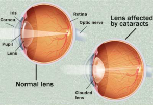
It’s a true statement that your grandparents’ cataract surgery is not the same as what you can expect today from cataract surgery. Improved surgical techniques, as well as the use of, and upgrades to intraocular lenses (IOLs), has significantly changed ophthalmology within the last 75 years.
Cataract Surgery in the Early 1900s
In the first half of the twentieth century, cataract surgery meant the complete removal of the crystalline lens in the eye. Removing the lens left the patient aphakic (without the natural lens of the eye), and dependent upon very thick glasses, or hard contact lenses to be able to achieve some visual acuity. The procedure was done in a hospital, and large incisions, followed by stitches, were necessary for the surgery.
Winds of Change for Cataract Surgery
But things started changing following World War II and two significant events in the life of Dr. Harold Ridley, an eye surgeon, and pioneer in cataract surgery. First, as a surgeon, Ridley had treated a pilot in the war who had chunks of polymethylmethacrylate (PMMA) embedded in his eye following the shattering of his airplane cockpit canopy. Ridley noted that the pilot was suffering no long-term adverse effects. The second was the inquisitive nature of a medical student who was unaware that the lens was not replaced following the removal of a cataract. This question led Ridley to research other methods. The question raised by the medical student, and a belief that a material could be placed in the human body without adverse effects lead to the development of the first IOL.
Technology Changes Gain Momentum in Cataract Surgery
Early on, there was a high failure rate with these IOLs. Ridley continued to refine the surgery process, including the evolution to only remove the cortex (center) of the lens, therefore leaving the lens capsule intact and open for the insertion of the new lens. At first, everyone received the same power lens (20.0) and wore glasses with a prescription similar to that worn prior to surgery. With the development in the 1970s of A-scan ultrasound technology techniques for eye measurements, patients received a more correctly-powered lens, providing better visual outcomes than had been possible in the past.
Another important advancement was the introduction of phacoemulsification in the 1980s, (the process of breaking up the lens before removal). Before phacoemulsification it was necessary for the surgeon to create 6-7mm incisions in order to remove the cataract intact. With phacoemulsification allowing for the removal of small bits of the cataract at a time, incision size was reduced to just 3.5mm, leaving the patient with a small incision that would self-heal without stitches. Even the use of silicone material for the IOL’s meant that it could be rolled up and inserted into the capsule through the same small incision.
Even within the last few decades the advancements with IOL technology is changing the way patients see following cataract surgery. Stay for part two of this blog post which will highlight these advancements.
Source: Lasik Rapid City Website
http://www.lasikrapidcity.com/blog/detail/2010/03/04/the-evolution-of-the-intraocular-lens-in-cataract-surgery.html


